
MRI Integration With PACS and EMR Systems: Technical Requirements Guide
If you work in a hospital radiology department or manage healthcare IT infrastructure, you already know that getting MRI scanners, Picture Archiving and Communication Systems (PACS), and Electronic Medical Record (EMR) systems to talk to each other is far from plug-and-play. These are complex, data-heavy systems that carry sensitive patient information, and when they don't communicate properly, the consequences ripple across clinical workflows, diagnostic turnaround times, and patient outcomes.
This guide breaks down exactly what it takes to integrate MRI systems with PACS and EMR platforms in 2026, covering the protocols, network infrastructure, security requirements, and common integration pitfalls you need to know before your next deployment or upgrade.
Understanding the Three-System Architecture
Before getting into the technical requirements, it helps to be clear about what each system actually does and why all three need to work in sync.
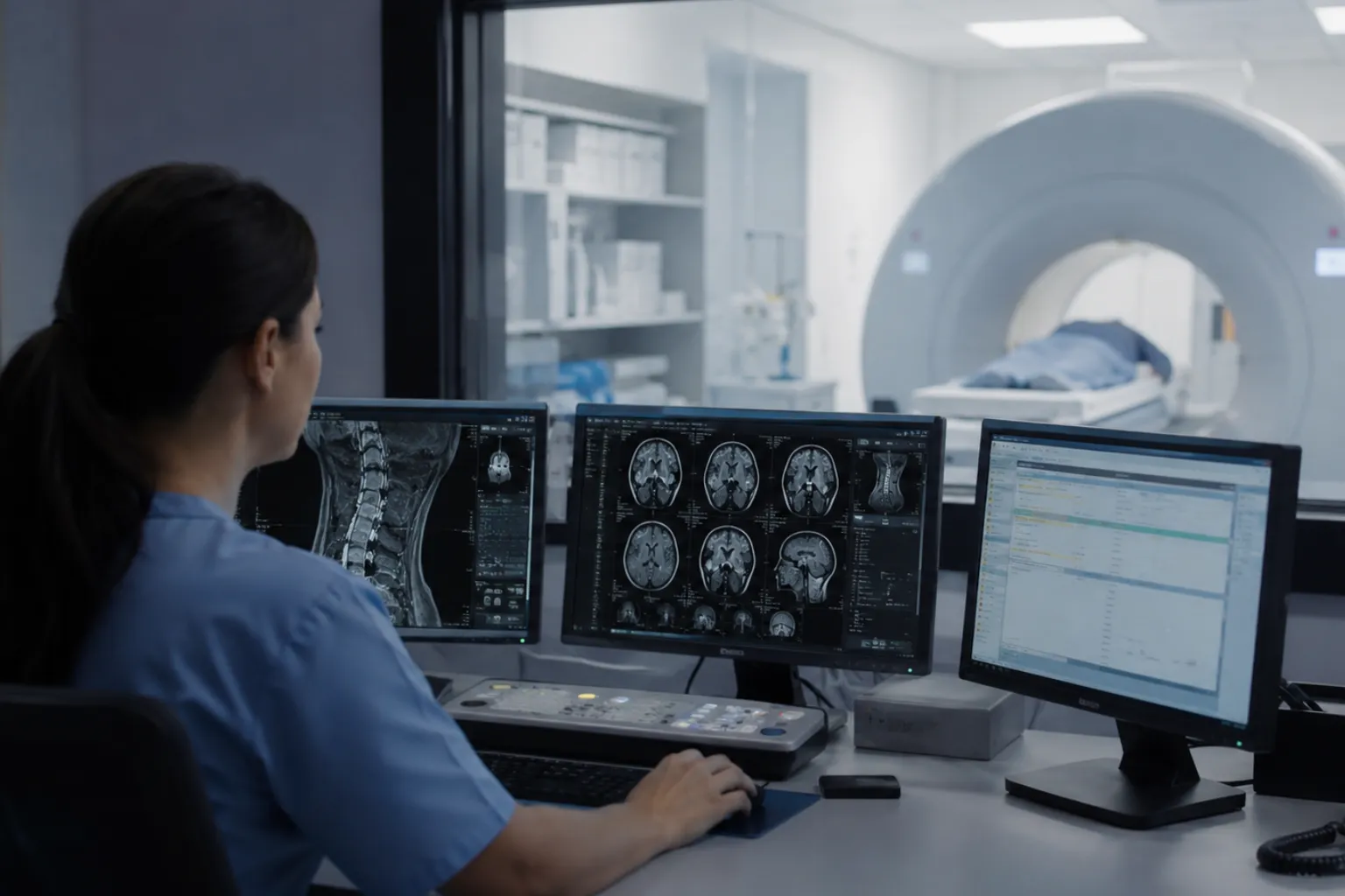
What Is PACS?
PACS (Picture Archiving and Communication System) is the digital backbone of medical imaging. It allows radiologists, technicians, and doctors to store, retrieve, manage, and share images like X-rays, CT scans, and MRIs efficiently. Modern PACS solutions go well beyond simple storage; they support 3D reconstruction, AI-assisted radiology, and real-time viewing from remote locations. Think of PACS as the central hub where your MRI scanner sends its output and where radiologists go to read those images. If you want a deeper breakdown of how PACS, EMR, and HIS systems relate to one another within a hospital setup, this guide to PACS, EMR, and HIS healthcare systems is a good place to start.
What Is an EMR?
An EMR (Electronic Medical Record) system is the organization-wide clinical record for a patient. It manages a patient's overall medical history within a specific practice or organization, including diagnoses, medications, allergies, immunizations, lab results, progress notes, and imaging reports. Unlike PACS, which is radiology-specific, the EMR is the broader clinical home for all patient data across departments.
Where Does MRI Fit In?
Your MRI scanner is the modality, meaning it generates the raw imaging data. Imaging equipment like CT, MRI, and ultrasound systems generate DICOM images and metadata that feed directly into PACS. But the ordering workflow starts in the EMR, the image interpretation happens in PACS, and the final diagnostic report needs to land back in the EMR so every treating clinician can access it. Keeping that loop tight is what integration is all about.
The Core Integration Protocols You Must Understand
There are three major standards that govern how MRI data moves across these systems. Getting fluent in all three is non-negotiable for any integration project in 2026.
DICOM: The Language of Medical Images
DICOM (Digital Imaging and Communications in Medicine) is the global standard for managing, storing, and transmitting medical images. DICOM ensures that images carry consistent metadata, including patient identifiers, imaging parameters, and timestamps, so files can be viewed, shared, and archived across different devices and platforms. You can review the full technical specification directly on the official DICOM Standard website.
Every MRI scan your system produces is a DICOM file. That file includes not just the pixel data of the image but also a structured header with the patient's name, study date, acquisition parameters, and a unique Study Instance UID. DICOM not only defines an interoperable data format for storing medical images but also includes metadata encapsulated in structured tags, ensuring compatibility across different systems and forming the backbone of digital imaging workflows in modern healthcare.
Modern MRI-PACS integration also uses DICOMweb, a RESTful API extension of the DICOM standard that allows web-native image access and retrieval. This is especially important for cloud-based PACS deployments and remote access scenarios that are now common in 2026.
HL7: Clinical Data Messaging
HL7 (Health Level Seven) manages the exchange of clinical and administrative data such as patient demographics, imaging orders, and reports. HL7 v2/v3 is widely used for structured message-based communication between systems.
In practical terms, HL7 messages carry the order from the EMR to PACS, the patient demographics that get pre-loaded onto the MRI scanner via the DICOM Modality Worklist (DMWL), and the finalized radiology report that gets pushed back into the EMR as a result message. Without solid HL7 integration, technologists end up manually entering patient data at the scanner console, which is slow and introduces transcription errors.
The canonical imaging pattern bridges HL7-based systems like the RIS with DICOM-based modalities and PACS by defining semantic mappings, then makes patient and order information available to acquisition via DICOM Modality Worklist (MWL).
FHIR: The Modern API Layer
FHIR (Fast Healthcare Interoperability Resources) is the newer web-based standard that uses APIs for real-time, flexible data sharing, especially useful for cloud-based PACS and mobile or remote access to imaging data.
If HL7 v2 is a fax machine and HL7 v3 is an early email client, FHIR is the modern REST API. It uses standard web technologies (JSON, XML, HTTP) to expose healthcare data as resources, making it far easier to integrate with third-party applications, mobile viewers, and AI analytics platforms. Key FHIR resources for imaging integration include ServiceRequest for the order, ImagingStudy to represent the DICOM study, and DiagnosticReport to package the finalized report.
In 2026, SMART on FHIR is also gaining ground as a way to launch contextual imaging viewers directly from within the EMR, letting a referring physician click on an imaging order and pull up the PACS viewer without a separate login.
Step-by-Step: How the MRI-PACS-EMR Workflow Actually Flows
Understanding the data flow is essential before configuring any integration. Here is the typical end-to-end sequence:
- Order Placement: A physician orders an MRI study through the EMR. That order is transmitted to the Radiology Information System (RIS) and PACS via an HL7 ORM (Order Request Message).
- Modality Worklist: The MRI scanner polls the PACS or RIS for scheduled studies using the DICOM Modality Worklist. Patient demographics, accession numbers, and study descriptions auto-populate on the scanner console, eliminating manual entry.
- Image Acquisition: The MRI scan is performed. The imaging device produces a DICOM file, which includes both the image and metadata, and this file is automatically transmitted to the PACS server for storage.
- Report Creation: The radiologist opens the study in PACS, reads the images using a diagnostic workstation, and dictates or types the report, often using a voice recognition system integrated with the PACS viewer.
- Result Delivery: The finalized ORU (Observation Result) message returns report status to the EHR, making it visible to the ordering physician and care team inside the EMR.
- Bidirectional Access: Radiologists can pull relevant clinical history directly from the EMR while viewing scans in PACS, and diagnostic reports created in PACS are automatically pushed into the patient's EMR.
Let’s Build Your Dream Hospital
Whether you’re planning a new hospital, expanding an existing facility, or upgrading your healthcare technology, Actiss Healthcare is here to guide you every step of the way. Let us help you turn your vision into reality. Contact us today for a free consultation & learn more about our services and how we can support your next healthcare project.

Network Infrastructure Requirements
Network performance is where MRI integration projects most often run into trouble. MRI studies are large. A single brain MRI with multiple sequences can run into several gigabytes, especially with high-resolution 3D acquisitions. Your network has to handle that volume reliably, every day, at peak scan times. It's worth noting that network cabling, switching hardware, and power distribution all tie back to your facility's MEP infrastructure - a subject covered in detail in this hospital MEP systems planning guide.
Bandwidth Specifications
| Network Segment | Minimum Recommended Speed | Notes |
|---|---|---|
| MRI Scanner to PACS (LAN) | 1 Gbps (Gigabit Ethernet) | Higher-field MRI (3T, 7T) generates larger datasets; 10 Gbps preferred |
| PACS to Diagnostic Workstations (LAN) | 1 Gbps | Radiologist workstations must render images without perceptible lag |
| PACS to EMR (LAN/WAN) | 100 Mbps minimum | Report text is small; image thumbnails and links require more |
| Remote/Teleradiology (WAN) | High-bandwidth WAN, 50 Mbps+ | VPN encryption adds overhead; latency under 50ms preferred |
| Cloud PACS Access | Fiber broadband with redundant ISP | Content Delivery Networks (CDNs) help reduce latency |
A properly configured network should have failover mechanisms, backup internet connections, and automatic rerouting to ensure uninterrupted access to data, because even a few seconds of lag can frustrate users and reduce adoption.
LAN vs. WAN Considerations
Within the hospital or imaging center, a robust LAN, often Gigabit Ethernet or faster, is needed to transmit images from modalities to the PACS archive and from the archive to workstations. For teleradiology or multi-site enterprises, secure and high-bandwidth WAN connections are necessary to share images between locations or provide remote access.
Security and Compliance Requirements
Patient imaging data is among the most sensitive data a healthcare organization holds. Your integration architecture has to address security at every layer, not just at the perimeter.
Encryption Standards
Data encryption is a core requirement: TLS/SSL for data in transit, AES-256 for data at rest, and end-to-end encryption for wireless device communications. Any DICOM send operation over a network should use Secure DICOM (DICOM over TLS), and all HL7 or FHIR API traffic should be encrypted over HTTPS.
Access Control and Authentication
Role-based access control, multi-factor authentication, and comprehensive audit logs are needed to track who accessed or modified device data. In a well-configured PACS-EMR integration, a referring physician should only be able to view studies linked to their patients, while a radiologist can access a broader worklist. Integration engines and middleware should log every message transaction for audit trail purposes.
Regulatory Compliance
Different regions have different regulations: HIPAA in the U.S. requires rigorous data protection measures, while GDPR in the EU adds even stricter rules around patient consent and data exchange. Your integration project needs legal review against the applicable regulatory framework before go-live. The U.S. Department of Health and Human Services HIPAA resource center is a reliable reference point for understanding exactly what the law requires of covered entities and their business associates.
Integrated systems must comply with healthcare data privacy regulations such as HIPAA and GDPR, ensuring encryption, user authentication, audit logs, and secure transmission of imaging and patient data. Compliance with HIPAA, HITECH, and GDPR standards ensures secure handling of medical images and records across all stages of storage and transmission. If your team needs expert support in mapping these compliance requirements to your specific technology setup, healthcare technology consultancy services can help you structure the right compliance architecture from the ground up.
Physical and Network Security
The network must be highly secure, incorporating firewalls, intrusion detection systems, and secure protocols like VPNs for remote access to protect against unauthorized access and cyber threats. Don't overlook physical security either: MRI scanner rooms, PACS server areas, and diagnostic workstations all need physical access controls.
Middleware and Integration Engines
In most real-world deployments, you can't connect an MRI scanner directly to an EMR without something in between. That's where middleware and integration engines come in.
What Integration Engines Do
Healthcare providers can connect disparate systems like EHR/EMR, PACS, RIS, or HIS into a single framework and centralized messaging hub for sharing enterprise imaging-related data. All connections to and from a PACS, vendor neutral archive (VNA), and modalities can be managed through a single workflow engine supporting DICOM, DICOMweb, HL7, or FHIR, either onsite or remotely.
Middleware solutions translate and route data between DICOM and HL7 systems, ensuring that data flows across different platforms and applications without conflict. A good integration engine is vendor-agnostic, meaning it can connect systems from different manufacturers and different generations without requiring you to replace everything at once.
HL7 vs. FHIR: Choosing the Right Layer
| Feature | HL7 v2 | FHIR R4/R5 |
|---|---|---|
| Data Format | Pipe-delimited text messages | JSON or XML over REST APIs |
| Implementation Complexity | Moderate (requires interface engines) | Lower for modern systems with standard web tools |
| Real-Time Data Sharing | Batch or near-real-time | True real-time via API calls |
| Legacy System Support | Very high | Limited without adapters |
| Mobile/Web App Integration | Poor | Excellent (native web standard) |
| MRI-Specific Imaging Resource | ORM/ORU message segments | ImagingStudy, DiagnosticReport resources |
In 2026, most major EMR vendors like Epic, Oracle Health (formerly Cerner), and Meditech support both HL7 v2 and FHIR interfaces. If you're building a new integration from scratch, lead with FHIR but keep HL7 v2 compatibility for the legacy systems that aren't going away anytime soon.
Common Integration Challenges and How to Fix Them
Vendor Compatibility Issues
One of the most persistent challenges in integration is compatibility. Not all PACS or EMR systems are designed to speak the same language. Some use proprietary data formats or outdated protocols, which makes sharing information complex. Even when systems use standards like DICOM or HL7, different interpretations of those standards can still create barriers.
The fix: Conduct a system inventory and compatibility audit before integration begins, use middleware that can translate and mediate between disparate systems, and insist on vendors supporting interoperability standards like FHIR and DICOMweb.
Patient Identity Mismatch
One of the most serious data quality problems in MRI-PACS-EMR integration is mismatched patient identifiers. If the patient's medical record number (MRN) is formatted differently in the EMR versus the PACS, studies can get linked to the wrong patient. You need a Master Patient Index (MPI) or an enterprise-level patient matching strategy as part of your integration design.
Legacy System Limitations
Older PACS or EHR platforms often lack support for modern interoperability standards like DICOMweb, HL7 FHIR, or RESTful APIs. Replacing these systems entirely can be costly and disruptive. The practical path for most organizations is smart integration adapters or shim layers that bridge older systems to newer protocols without full replacement.
Latency and Performance Degradation
Even with adequate bandwidth, poor network configuration can cause significant slowdowns. Content Delivery Networks (CDNs), edge caching for frequently accessed prior studies, and low-latency switching hardware all contribute to the kind of sub-second image loading that radiologists expect on diagnostic workstations.
Storage Architecture for MRI Data
MRI data puts serious pressure on your storage infrastructure. A single high-field MRI study with multiple sequences can be 500 MB to 2 GB or more. At a busy imaging center performing 50 to 100 MRI studies per day, that adds up fast.
Tiered Storage Strategy
- Hot Storage (SSD/NVMe): Recent studies, typically within 30 to 90 days, should be on fast local storage that PACS can serve to workstations with minimal latency.
- Warm Storage (SAN/NAS with RAID): Studies from 90 days to 2 years go here. Fast enough for clinical retrieval but lower cost per TB.
- Cold/Archive Storage (Cloud or Tape): PACS maintains long-term archives for legal, research, and clinical reference purposes. In the U.S., most states require diagnostic images to be retained for a minimum of 7 to 10 years for adults.
Vendor Neutral Archives (VNAs)
A Vendor Neutral Archive stores DICOM images in a standardized format independent of any specific PACS vendor. This protects you from vendor lock-in and makes it much easier to migrate between PACS platforms in the future, a major concern for hospital IT teams planning 10-year infrastructure lifecycles.
AI and Emerging Integrations in 2026
The integration of AI tools into the MRI-PACS-EMR pipeline is no longer experimental. AI-powered analytics are now being used to prioritize critical cases, with outcomes showing turnaround time for critical scans dropping from 4 hours to under 60 minutes in integrated radiology departments. These AI tools typically plug into PACS via DICOM or DICOMweb interfaces and feed their outputs, like a priority flag or a preliminary finding, back into the structured report workflow. To understand how AI, IoT, and automation are being designed into hospital infrastructure more broadly, see this detailed write-up on smart hospitals, IoT, and AI automation in healthcare design.
For AI to work well in an integrated environment, the integration architecture needs to support bi-directional communication: the AI model receives the DICOM study, processes it, and writes its results back into the PACS or EMR as a structured annotation or alert. This requires solid API design and well-defined data governance policies from the start.
Pre-Integration Checklist
Before you kick off any MRI-PACS-EMR integration project, work through this checklist to avoid the most common failure points:
- Complete a full system inventory of all MRI scanners, PACS versions, and EMR platforms in scope
- Audit DICOM conformance statements for every scanner model to confirm compatibility
- Map all HL7 message types currently in use (ADT, ORM, ORU, SIU) across existing systems
- Identify patient identifier formats across systems and plan MPI strategy
- Assess current network bandwidth utilization and identify upgrade needs
- Review all HIPAA/GDPR obligations with legal and compliance teams
- Define a testing and validation protocol before go-live
- Establish downtime procedures and fallback workflows
- Train radiologist and clinical staff on new integrated workflows
- Plan post-go-live monitoring with defined KPIs (report turnaround time, error rates, image retrieval speed)
Conclusion
MRI integration with PACS and EMR systems in 2026 is technically achievable for any organization willing to invest the right time in planning and infrastructure. The standards are mature, DICOM handles the images, HL7 handles the clinical messaging, and FHIR bridges the gap for modern API-based communication. The real work is in the details: network design, patient identity management, security architecture, and thorough pre-go-live testing. Projects of this complexity benefit greatly from structured oversight - the kind that a dedicated hospital project management consultancy brings to high-stakes technology deployments. When this integration runs well, radiologists get clinical context at the point of image reading, referring physicians get reports inside their EMR workflow without extra clicks, and patients get faster diagnoses. That outcome is absolutely worth the technical effort it takes to get there.
Let’s Build Your Dream Hospital
Whether you’re planning a new hospital, expanding an existing facility, or upgrading your healthcare technology, Actiss Healthcare is here to guide you every step of the way. Let us help you turn your vision into reality. Contact us today for a free consultation & learn more about our services and how we can support your next healthcare project.
FAQs
1. What is the difference between DICOM and HL7 in MRI-PACS-EMR integration?
DICOM is the standard specifically designed for medical images. It governs how MRI scan data, along with its metadata like patient ID and scan parameters, is formatted, transmitted, and stored between the scanner and PACS. HL7 handles the clinical and administrative text data, like imaging orders, patient demographics, and finalized radiology reports, that flow between the EMR, RIS, and PACS. Both protocols work together in a fully integrated radiology environment, with HL7 carrying the order workflow and DICOM carrying the actual image data.
2. How much network bandwidth does an MRI-PACS integration actually need?
At a minimum, the local network segment between your MRI scanner and PACS should run on Gigabit Ethernet (1 Gbps). For high-field 3T or 7T MRI systems that generate very large datasets or for departments running multiple scanners simultaneously, 10 Gbps connections are strongly preferred. For remote reading or teleradiology, a dedicated high-bandwidth WAN link with a redundant internet connection and latency under 50 milliseconds is the practical standard.
3. Can older PACS systems integrate with modern EMR platforms like Epic or Oracle Health?
Yes, but it typically requires middleware or an integration engine to bridge the gap. Older PACS systems often rely on HL7 v2 message-based communication and may not support FHIR APIs or DICOMweb natively. Integration adapters can translate between older protocols and newer standards, allowing legacy PACS to exchange data with modern EMR platforms without requiring full system replacement. That said, legacy systems may limit the depth of bidirectional data exchange, so a full upgrade roadmap is usually still advisable.
4. What are the HIPAA compliance requirements specific to MRI-PACS-EMR data exchange?
Under HIPAA, all protected health information (PHI) transmitted between MRI, PACS, and EMR systems must be encrypted in transit (using TLS/SSL) and at rest (using AES-256 or equivalent). Systems must implement role-based access controls, maintain complete audit logs of who accessed or modified patient data, and have documented breach notification procedures. Any third-party middleware vendor or cloud PACS provider also needs to operate under a signed Business Associate Agreement (BAA) with your organization.
5. What is a Vendor Neutral Archive (VNA) and does my facility need one?
A Vendor Neutral Archive is a storage system that holds DICOM images in a standardized format that isn't tied to any specific PACS vendor. If you ever need to switch PACS vendors, a VNA lets you migrate without losing access to years of historical MRI studies. For large health systems, academic medical centers, or any facility planning a PACS upgrade in the next five to ten years, a VNA is a very practical investment. It also makes it much easier to share imaging data across multiple facilities or with external specialists, since the data lives in a format any standards-compliant viewer can read.